2MSB

STRUCTURE OF A C-TYPE MANNOSE-BINDING PROTEIN COMPLEXED WITH AN OLIGOSACCHARIDE


Experimental Data Snapshot

  • Method: X-RAY DIFFRACTION
  • Resolution: 1.70 Å
  • R-Value Work: 0.174 
  • R-Value Observed: 0.174 

wwPDB Validation   3D Report Full Report


This is version 2.0 of the entry. See complete history


Literature

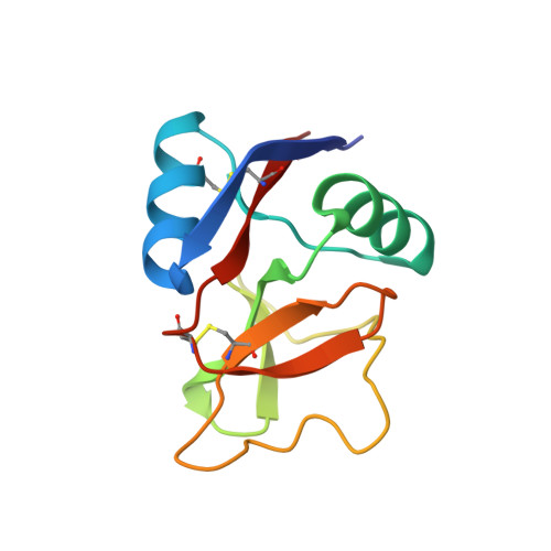
Structure of a C-type mannose-binding protein complexed with an oligosaccharide.

Weis, W.I.Drickamer, K.Hendrickson, W.A.

(1992) Nature 360: 127-134

  • DOI: https://doi.org/10.1038/360127a0
  • Primary Citation of Related Structures:  
    2MSB

  • PubMed Abstract: 

    C-type (Ca(2+)-dependent) animal lectins such as mannose-binding proteins mediate many cell-surface carbohydrate-recognition events. The crystal structure at 1.7 A resolution of the carbohydrate-recognition domain of rat mannose-binding protein complexed with an oligomannose asparaginyl-oligosaccharide reveals that Ca2+ forms coordination bonds with the carbohydrate ligand. Carbohydrate specificity is determined by a network of coordination and hydrogen bonds that stabilizes the ternary complex of protein, Ca2+ and sugar. Two branches of the oligosaccharide crosslink neighbouring carbohydrate-recognition domains in the crystal, enabling multivalent binding to a single oligosaccharide chain to be visualized directly.


  • Organizational Affiliation

    Department of Biochemistry and Molecular Biophysics, Columbia University, New York, New York 10032.


Macromolecules
Find similar proteins by:  (by identity cutoff)  |  3D Structure
Entity ID: 1
MoleculeChains Sequence LengthOrganismDetailsImage
MANNOSE-BINDING PROTEIN-A
A, B
115Rattus rattusMutation(s): 0 
UniProt
Find proteins for P19999 (Rattus norvegicus)
Explore P19999 
Go to UniProtKB:  P19999
Entity Groups  
Sequence Clusters30% Identity50% Identity70% Identity90% Identity95% Identity100% Identity
UniProt GroupP19999
Sequence Annotations
Expand
  • Reference Sequence
Oligosaccharides

Help

Entity ID: 2
MoleculeChains Length2D Diagram Glycosylation3D Interactions
alpha-D-mannopyranose-(1-2)-alpha-D-mannopyranose-(1-3)-[alpha-D-mannopyranose-(1-3)-[alpha-D-mannopyranose-(1-6)]alpha-D-mannopyranose-(1-6)]beta-D-mannopyranose-(1-4)-2-acetamido-2-deoxy-beta-D-glucopyranose
C
7N/A
Glycosylation Resources
GlyTouCan:  G30704AF
GlyCosmos:  G30704AF
GlyGen:  G30704AF
Experimental Data & Validation

Experimental Data

  • Method: X-RAY DIFFRACTION
  • Resolution: 1.70 Å
  • R-Value Work: 0.174 
  • R-Value Observed: 0.174 
  • Space Group: P 1 21 1
Unit Cell:
Length ( Å )Angle ( ˚ )
a = 34.81α = 90
b = 85.16β = 97.41
c = 39.55γ = 90
Software Package:
Software NamePurpose
X-PLORmodel building
X-PLORrefinement
X-PLORphasing

Structure Validation

View Full Validation Report


Entry History 

Deposition Data

Revision History  (Full details and data files)

  • Version 1.0: 1993-10-31
    Type: Initial release
  • Version 1.1: 2008-03-03
    Changes: Version format compliance
  • Version 1.2: 2011-07-13
    Changes: Non-polymer description, Version format compliance
  • Version 2.0: 2020-07-29
    Type: Remediation
    Reason: Carbohydrate remediation
    Changes: Advisory, Atomic model, Data collection, Derived calculations, Other, Structure summary